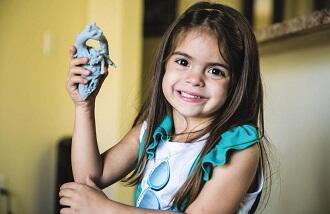
At Nicklaus Children’s Hospital in Miami, director of pediatric cardiovascular surgery Redmond Burke dedicates his intellect and his hands to repairing tiny hearts. Burke performs challenging operations, often for children who have nowhere else to turn. In the complex yet highly tactile task of rebuilding the vital organ, he has a new ally: 3D printing.
Mia
For patients with rare defects, Burke must plan procedures based on each child’s condition and anatomy. In the case of Mia Gonzalez, that meant untangling a double aortic arch, a structural defect in which a complete vascular ring wraps around the trachea or esophagus, restricting airflow and causing coughing and frequent respiratory infections. Before coming to Nicklaus Children’s Hospital, Mia spent the first four years of her life in and out of hospitals, misdiagnosed with asthma and struggling to breathe and swallow.
Using Stratasys solutions, the hospital created an anatomically precise 3D model of Mia’s heart, directly from her CT scan. With the model, Burke and his team were able to figure out which part of her arch should be divided to achieve the best physiological result. The clearer plan that resulted from the model reduced operating-room time by two hours, according to the hospital - significant in terms of risk to the patient as well as cost.
“We used
the most sophisticated imaging systems, echocardiography and CT angiography, to
study Mia’s heart”, Burke said. “But for a surgeon, there was something more
compelling about holding an exact replica of her heart in my hands. My team
could visualize the operation before we started. We knew the safest approach,
and confidently made a smaller incision. I’ve seen surgeons get lost doing rare
operations like Mia’s. The 3D model allowed me to proceed through Mia’s
operation with confidence because I knew her unique anatomy perfectly”.
He also used it to prepare Mia’s family for the surgery. He showed it to them, and said: “This is what’s choking your baby. This is your baby’s heart, and this is how I’m going to repair it”. “From having four and a half years of not knowing, to all of the sudden in less than a two-month timeframe, she’s back out of her surgery and back to normal”, said Katherine Gonzalez, Mia’s mother, “that’s been a great experience for us”.
Adenelie
Adenelie Gonzalez was born with a lethal heart defect called a total anomalous pulmonary venous connection. Previous surgeries she underwent as a newborn and at nine months, and four catheterizations, provided only temporary help. By age four she weighed only 28 pounds, and her health was deteriorating rapidly. Her cardiologist had difficulty finding a surgeon willing to take on a high-risk surgery.
“Looking at Adenelie’s X-rays and catheterizations, I thought she was inoperable”, said Burke. “Her deformity was one-of-a-kind”. Images on a computer screen were not enough. “But I thought that holding and manipulating a flexible 3D replica of this child’s heart might allow me to design an operation that hadn’t been done before. We could configure the necessary patches to create the exact shapes and dimension to match her deformed pulmonary veins”.
The challenge was creating a 3D model with approximately the same amount of flexibility as the human heart. Chelsea Balli, cardiac surgery biomedical engineer at Nicklaus Children’s Hospital, determined that a material with a Shore A value of 60 would closely match the properties of the heart. A Connex3 3D Printer from Stratasys, which combines photopolymers for a range of characteristics, achieved the right feel.
Thinking by feeling
Burke carried the model heart in his gym bag, and when he had free minutes he would reach into the bag, feel the model, look at it, flip it over, and run through possible reconstructions in his mind. He manipulated the model as he later would Adenelie’s living heart, moving vessels around to explore possible repairs. In one of those idle moments he figured out a practical solution.
Burke determined the exact size and geometry needed for the repair parts so they could be prepared in advance, minimizing the time Adenelie would have to spend on bypass. Later, he used the model to explain the surgery to Adenelie’s parents, and to prep his team.
In the operating room, Adenelie’s heart was connected to a bypass machine and cooled to freezing temperatures so it could be manipulated without damage. Burke made the repairs and her heart was rewarmed. It began beating normally. For the first time in her life, Adenelie’s internal heart pressures measured normal.
“Without a 3D printed model, I might not have been able to figure out the repair method that I used, and I’m not sure if the operation would have been successful”, Burke said.


